title:: Patient-Specific Modeling and Quantification of the Aortic and Mitral Valves From 4-D Cardiac CT and TEE
publisher:: IEEE
people:: Razvan Ionasec, Ingmar Voigt, Bogdan Georgescu, Yang Wang, Helene Houle, Fernando Vega-Higuera, Nassir Navab, Dorin Comaniciu
organization:: Siemens Corporate Research, Siemens Healthineers, Technical University of Munich, Friedrich-Alexander-Universität Erlangen-Nürnberg
domain:: Medical Imaging, Machine Learning, Computer Vision, Cardiology
link:: https://doi.org/10.1109/tmi.2010.2048756
citations:: 194 (OpenAlex) · 211 (Semantic Scholar) — as of 14 October 2026
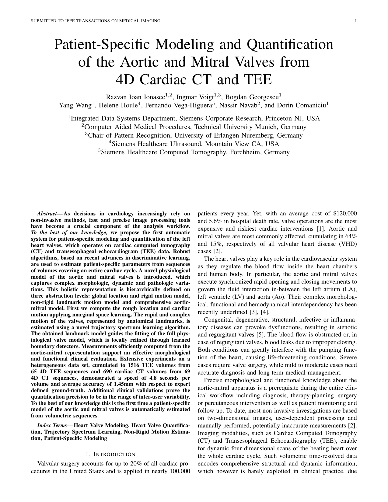
First page of the paper — author copy hosted by the FAU Pattern Recognition Lab. Corresponding-author contact details redacted.
Summary
The first automatic system for patient-specific modelling and quantification of the left heart valves operating on both cardiac CT and transesophageal echocardiography. The paper introduces a physiological model of the aortic and mitral valves that captures complex morphologic, dynamic and pathologic variation, defined hierarchically on three abstraction levels: a global location and rigid motion model, a nonrigid landmark motion model, and a comprehensive aortic-mitral model.
Parameters are estimated from volume sequences covering an entire cardiac cycle using discriminative learning. Rough location and cardiac motion come from marginal space learning; the rapid and complex motion of the valves, represented by anatomical landmarks, is estimated with a trajectory spectrum learning algorithm; the resulting landmark model then guides the fitting of the full physiological valve model, locally refined by learned boundary detectors. Measurements computed from the aortic-mitral representation support morphological and functional clinical evaluation.
Data Points
- First patient-specific model of the aortic and mitral valves automatically estimated from volumetric sequences
- Validated on a heterogeneous dataset: 1,516 TEE volumes from 65 4-D TEE sequences and 690 CT volumes from 69 4-D CT sequences
- 4.8 seconds per volume; average accuracy 1.45 mm against expert-defined ground truth
- Quantification precision shown to lie within the range of inter-user variability
- Three-level hierarchy: global rigid motion (marginal space learning), nonrigid landmark motion (trajectory spectrum learning), full valve model (learned boundary detectors)
- The method line behind Siemens Healthineers heart-valve software and the Thomas Edison Patent Award for System for Planning Heart Valve Surgery
Reference
IEEE Transactions on Medical Imaging 29(9):1636-1651, 2010. doi:10.1109/tmi.2010.2048756 · PubMed 20442044
194-211 citations (OpenAlex / Semantic Scholar, October 2026) - my most-cited work, first author
Part of the complete record in Scientific Publications - Peer-Reviewed Research Record.